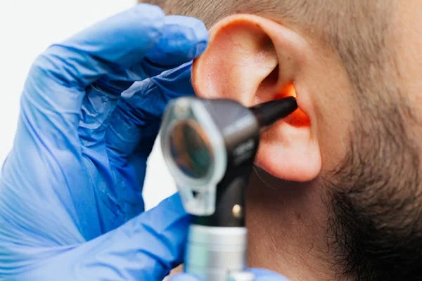
In the ever-evolving healthcare industry, technological innovations are increasingly significant in improving diagnosis, treatment, and patient outcomes. One of the latest technological advancements is using artificial intelligence (AI) in otoscope image analysis. Otoscopes are widely used medical instruments for examining the ear, enabling healthcare professionals to detect ear infections, earwax buildup, and eardrum perforations. However, introducing AI-powered applications has transformed traditional otoscopy by automating image analysis, increasing diagnostic accuracy, and streamlining the overall process.
ai-powered applications for otoscope image analysis provide healthcare professionals with real-time insights and a more precise understanding of ear health conditions, ultimately leading to more effective treatments. This article will explore how AI is revolutionizing the analysis of otoscope images, how it works, and the benefits it offers to healthcare professionals and patients. Additionally, we will examine the prospects, potential challenges such as data privacy and security, and ethical considerations of integrating AI into otoscope technology.
What is an Otoscope?
Healthcare providers use otoscopes to check for signs of ear infections, foreign objects, earwax buildup, perforated eardrums, and other conditions that affect ear health. Traditionally, otoscopes were operated manually, with healthcare professionals visually inspecting the images displayed on the device. However, AI-powered otoscopes now make it possible to automate the process of image analysis, allowing healthcare professionals to focus on interpreting the results and making treatment decisions, rather than spending time on the initial visual inspection.
How AI Enhances Otoscope Image Analysis
AI, specifically machine learning (ML) and deep learning (DL), are the core technologies driving the revolution in otoscope image analysis. These technologies can recognize patterns, anomalies, and potential health concerns that might be challenging for human clinicians to detect in traditional settings.
Key Features of AI in Otoscope Image Analysis:
- Automated Image Processing: AI algorithms can process and interpret images faster than the human eye, automatically detecting abnormal patterns and conditions within ear images.
- Pattern Recognition: AI can identify specific visual patterns associated with various ear conditions, such as fluid in the middle ear (otitis media), perforated eardrums, and earwax buildup.
- Real-Time Feedback: AI-powered otoscopes provide healthcare providers instant feedback, helping them make quick and accurate decisions on diagnosis and treatment.
- Predictive Analysis: By analyzing a large dataset of historical ear health images, AI can predict potential future conditions, allowing for early intervention and better preventive care.
Applications of AI in Otoscope Image Analysis
AI-powered applications in otoscope image analysis have numerous practical applications in medical settings. These applications help healthcare professionals detect conditions more accurately and enable better patient care and faster diagnosis. Some of the most significant applications include:
Detection of Ear Infections
Ear infections are among the most common reasons patients visit healthcare providers. AI-powered otoscopes can detect signs of ear infections, including fluid accumulation, redness, and eardrum inflammation. By analyzing the ear images, the AI algorithm can highlight areas of concern and provide actionable insights, offering patients reassurance through improved early detection and treatment outcomes.
Earwax Detection and Removal
Excess earwax buildup can cause discomfort, hearing loss, and even infections. AI can help identify the severity of earwax accumulation by analyzing images of the ear canal. This allows healthcare providers to determine whether earwax removal is necessary and plan an appropriate action. AI’s ability to detect earwax early can prevent complications such as ear infections and hearing loss.
Chronic Ear Condition Monitoring
Chronic ear conditions, such as otitis media with effusion, require ongoing monitoring. AI-powered otoscope image analysis can track changes in the ear over time, enabling healthcare providers to detect new issues early and adjust treatment plans as needed. AI can also help identify patterns and trends in chronic conditions, providing valuable insights into the patient’s overall ear health.
Perforated Eardrum Detection
A perforated eardrum can lead to hearing loss and increase the risk of infections. AI systems are trained to analyze images of the eardrum to identify perforations or tears. Early detection of eardrum perforations can help prevent further complications and ensure timely medical intervention.
Detection of Tumors and Abnormal Growths
Although rare, tumours or abnormal growths can develop in the ear canal or middle ear. AI-powered image analysis can assist in detecting irregular growths that may not be immediately visible to the human eye. Early identification of such conditions is vital for initiating treatment and preventing further health issues.
How Does AI-Powered Otoscope Image Analysis Work?
AI-powered otoscope image analysis uses machine learning and deep learning techniques to process and interpret ear images. These systems are trained on large datasets of labelled images, enabling the AI to learn and identify various ear conditions with high accuracy. Let’s break down the technology behind these systems:
Machine Learning (ML) and Deep Learning (DL)
ML and DL are subsets of artificial intelligence that allow algorithms to learn and improve over time. In otoscope image analysis, machine learning models are trained on thousands of labelled images of healthy and unhealthy ears. As the model processes more data, it becomes better at identifying specific ear conditions. Deep learning algorithms, especially convolutional neural networks (CNNs), are particularly effective in analyzing images as they can learn intricate features within visual data, such as textures and patterns.
Computer Vision
Computer vision is the technology that enables machines to interpret and understand visual data. In the case of AI-powered otoscope image analysis, computer vision algorithms extract crucial visual information from otoscope images and identify potential issues such as inflammation, foreign objects, or damage to the eardrum. Computer vision is critical for providing accurate and actionable results.
Data Annotation
For AI systems to make accurate predictions, experts must carefully annotate datasets. This involves labelling ear images with information about specific conditions, such as ear infections, earwax buildup, or eardrum perforations. Once the AI model is trained on a large annotated dataset, it can analyze new otoscope images to detect similar conditions.
Advantages of AI-Powered Otoscope Image Analysis
AI-powered otoscope image analysis offers several key benefits to both healthcare providers and patients:
Enhanced Diagnostic Accuracy
AI models are trained to detect subtle patterns in images that may not be visible to the naked eye. This improves diagnostic accuracy and reduces the chances of misdiagnosis.
Time Efficiency
AI-powered systems process images rapidly, allowing healthcare providers to receive real-time results. This leads to faster diagnoses and reduces patient waiting times.
Consistency in Results
Unlike human examiners, AI-powered systems deliver consistent results each time, eliminating the risk of errors due to fatigue or human oversight.
Better Patient Outcomes
By detecting ear conditions early, AI enables healthcare providers to intervene promptly, preventing complications and improving patient outcomes. Early intervention can reduce the need for extensive treatments and ensure faster recovery.
Increased Access to Care
AI-powered otoscope image analysis, especially when integrated into telemedicine platforms, can increase access to ear health care, particularly in remote or underserved areas. Patients can receive accurate diagnoses from the comfort of their homes, with healthcare providers offering remote consultations.
The Future of AI in Otoscope Image Analysis
The future of ai-powered applications for otoscope image analysis holds significant promise. AI technology is expected to offer even more precise and comprehensive diagnostic capabilities as it advances. Some potential developments include:
Integration with Telemedicine
AI-powered otoscope image analysis can be integrated into telemedicine systems, allowing patients to use handheld otoscopes at home and send images to healthcare providers for analysis. This would provide more accessible and efficient ear care for patients in remote areas.
Personalized Healthcare
AI’s ability to process vast amounts of data can be used to personalize healthcare plans. By analyzing an individual’s medical history, AI can help create tailored treatment plans considering their specific ear health needs.
Augmented Reality (AR) and AI
Combining AI with augmented reality (AR) could provide healthcare professionals with enhanced visualizations of ear conditions during real-time diagnostics. AR could overlay relevant information on the otoscope image, such as potential diagnoses, providing doctors with additional insights.
Challenges and Considerations
Despite the many advantages of AI-powered otoscope image analysis, there are several challenges to consider:
1. Data Privacy and Security
Healthcare data is highly sensitive, and AI-powered systems must adhere to strict privacy regulations, such as HIPAA, to ensure patient confidentiality. Ensuring that AI systems are secure and patient data is protected is critical for widespread adoption.
2. Integration with Existing Systems
Integrating AI-powered otoscopes into existing healthcare infrastructure, such as electronic health records (EHRs), can be complex. AI systems must be compatible with existing technologies to ensure smooth operations.
3. Regulatory Hurdles
AI-powered medical devices must undergo rigorous testing and regulatory approval before being used in clinical practice. The approval process can be lengthy, but ensuring that the technology meets safety and efficacy standards is essential.
Conclusion
Ai-powered applications for otoscope image analysis are transforming how ear conditions are diagnosed and treated. With increased diagnostic accuracy, speed, and consistency, these applications improve patient outcomes and streamline the diagnostic process. The potential benefits of AI in otoscope image analysis extend to early detection, remote consultations, and personalized healthcare.
As technology advances, AI-powered otoscopes will likely become essential in ear health care, benefiting both healthcare providers and patients. However, data privacy, integration, and regulatory approval challenges must be addressed to ensure that AI in otoscope image analysis reaches its full potential.


